


Volume -
3 , - : 149-155 |
Back to browse issues page



Download citation:
BibTeX | RIS | EndNote | Medlars | ProCite | Reference Manager | RefWorks
Send citation to:


BibTeX | RIS | EndNote | Medlars | ProCite | Reference Manager | RefWorks
Send citation to:
Cytochrome P450 2C19 Deficiency as the Sole Predictor of Major Adverse Cardiovascular Events in Post-COVID-19 Patients. 3 https://doi.org/10.58209/ijwph.18.2.149
URL: http://daneshafarand.org/article-1-85701-en.html
URL: http://daneshafarand.org/article-1-85701-en.html
Keywords: CYP2C19, Cytochrome P450, Post-COVID-19, Major Adverse Cardiovascular Events (MACE), Myocardial Infarction, IL-6
| Abstract (HTML) (268 Views)
Full-Text: (46 Views)
Introduction
Infection with SARS-CoV-2 leads to a multisystem illness that, among other effects, can cause long-lasting changes in the heart after the infection has resolved. Researchers who have studied very large groups of people have found that those who have had COVID-19 are at a much higher risk of developing new heart disease within a year after they recover, such as heart attack, stroke, heart failure, and blood clots, even if they did not have any heart problems before [1, 2]. This set of heart-related problems after the illness, along with many other symptoms, makes up what have been termed Post-Acute Sequelae of SARS-CoV-2 infection (PASC), or Long COVID, and it is estimated that 10 to 30% of those who have had COVID-19 are dealing with it [3, 4].
A variety of pathobiological processes contribute to sustained cardiovascular risk in post-COVID-19. These include persistently impaired endothelial function, overactive platelets, microvascular thromboinflammation, autonomic dysregulation, and continuous high levels of inflammatory cytokines [5, 6]. However, an important component of the risk puzzle—one that has received little attention so far—is how the cytokine environment after COVID-19 influences the hepatic cytochrome P450 (CYP) enzyme system, the main metabolic system in the body, which, among other functions, handles the processing of both externally and internally produced vasoactive substances.
Cytochrome P450 2C19 (CYP2C19) is considered one of the most crucial CYP isoforms clinically in the field of cardiovascular medication. It is the enzyme that, in the liver, converts the antiplatelet prodrug clopidogrel into its active thiol metabolite [7]. It also metabolizes arachidonic acid into epoxyeicosatrienoic acids (EETs)—which are potent vasodilators and have antithrombotic properties [8]—and it is involved in the biotransformation of several endogenous vasoactive substances, including thromboxane precursors. Besides its pharmacological function, CYP2C19-derived EETs also directly protect the vascular endothelium by blocking NF-B–triggered inflammation, reducing leukocyte adhesion, and maintaining the balance of fibrinolysis [9, 10].
Significant transcriptional downregulation of CYP2C19 occurs during systemic inflammation. Pro-inflammatory cytokines IL-6 and TNF—both of which are present at significantly high levels during and after COVID-19 infection—lead to the activation of STAT3 and NF-B signaling pathways within hepatocytes, which in turn causes phosphorylation-dependent inhibition of hepatocyte nuclear factor 4 (HNF4), the main transcriptional regulator of CYP2C19 [11, 12]. At the same time, a continuous oxidative stress environment resulting from COVID-19-induced NADPH oxidase activation will cause post-translational heme degradation of the CYP2C19 protein via formation of the compound I ferryl intermediate [13]. Therefore, the combined transcriptional and post-translational repression of CYP2C19 by the post-COVID-19 inflammatory milieu provides a fully plausible biological explanation for the progressive enzymatic deficiency that can persist for weeks to months after viral elimination.
Although this mechanistic plausibility cannot be ruled out, to date, no study has prospectively quantified CYP2C19 enzymatic activity in post-COVID-19 patients and simultaneously assessed the relationship between CYP2C19 enzymatic activity and major adverse cardiovascular events (MACEs) using a matched post-COVID-19 control design [14, 15]. With its demographic and environmental characteristics—including high ambient temperatures, dust-related oxidative stress exposure, and, in addition to the historically high COVID-19 case burden—this population is particularly informative for studying post-COVID-19 cardiometabolic susceptibility [16]. Decreased CYP2C19 enzymatic activity is an associated factor for cardiovascular risk, even after adjusting for well-known risk factors and inflammatory markers [17].
This study aimed to investigate whether CYP2C19 enzymatic activity decreases substantially in post-COVID-19 individuals who develop MACEs compared with those who remain event-free.
Instrument and Methods
This prospective, single-center, case-control study was conducted at the Maysan Center for Heart Diseases and Surgery, Maysan Health Directorate, Amarah, Maysan Province, Iraq, from October 2022 to November 2024. This Center is the main tertiary referral center for Maysan governorate, with a registered population of about 1.1 million. The hospital’s cardiology department manages the full spectrum of acute and chronic cardiovascular diseases in the region, including all post-COVID-19 cardiovascular cases referred from the province’s primary and secondary care centers.
Altogether, 210 adult participants were recruited from the outpatient cardiology and post-COVID-19 follow-up clinics of the Maysan Center for Heart Diseases and Surgery and divided into two groups. Both groups consisted of individuals with PCR-confirmed COVID-19 infections during the time of study enrollment.
The case group (n=105) included adults who developed a new MACE (objectively confirmed) within six months of COVID-19 recovery, defined as a heart attack, deep vein thrombosis, ischemic stroke, or pulmonary embolism. The diagnosis of heart attack was based on the Fourth Universal Definition criteria (cardiac troponin rise/fall plus symptoms or ECG changes indicative of myocardial ischemia). Deep vein thrombosis (DVT) was detected by Duplex Doppler ultrasound. Ischemic stroke was based on clinical assessment and CT/MRI imaging. Pulmonary embolism (PE) was diagnosed through CT pulmonary angiography [19].
The control group (n=105) comprised individuals who had recovered from COVID-19 and visited the same institutional follow-up clinic during the same calendar period as the cases. Controls were matched to cases by age, sex, and BMI (2.5kg/m). Control participants included not only those who completed a structured six-month cardiovascular surveillance program but also those who did not develop any MACE. Recruitment of controls was prospective and concurrent with case identification, ensuring temporal comparability of post-infection inflammatory status.
A matched post-COVID-19 versus post-COVID-19 design such as this one excludes the confounding effect of infection history and highlights enzymatic depletion as a primary factor underlying adverse cardiovascular outcomes in the post-COVID-19 population. All patients admitted to the cardiac hospital during the research who met the inclusion criteria were enrolled. Each eligible case was recorded individually. Consequently, the sample size was not formally determined, because all available cases during the research were included.
Participants were eligible only if they were adults (>18 years) of either gender with a documented history of PCR-confirmed COVID-19 (controls were eligible if they attended follow-up after recovery) and had no prior diagnosis of cardiovascular disease. Participants were also excluded if they had conditions known to alter CYP2C19 expression/activity independent of COVID-19 (e.g., cirrhosis/major liver dysfunction with ALT or AST>3× upper normal limit, chronic kidney disease with eGFR <45mL/min/1.73m², active cancer, or type 1 diabetes), current or recent (last 30 days) use of CYP2C19 inhibitors (e.g., fluconazole, fluvoxamine, ticlopidine, and chronic omeprazole) or potent CYP2C19 inducers (e.g., rifampicin, carbamazepine, St John’s Wort), confirmed/suspected hereditary thrombophilia (e.g., factor V Leiden, prothrombin G20210A, protein C/S or antithrombin III deficiency), active smoking (excluded due to smoking-induced effects on multiple CYP enzymes including CYP2C19) [20], pregnancy/lactation, or BMI>35kg/m².
All biochemical analyses were performed in the hospital’s accredited clinical biochemistry laboratory under standardized pre-analytical conditions, and the laboratory staff was not aware of the participants’ group allocations.
The study was conducted in accordance with the ethical standards set forth in the 1964 Declaration of Helsinki and its six amendments through 2013 [18]. Each potential subject was invited to a clear and detailed informed-consent discussion, in which the main points of the study were presented, the types and quantities of biological specimens were described, and it was explained that the data would be fully anonymized. In addition, the participants’ rights to withdraw at any time without their care being affected were explained in detail. Subsequently, written informed consent was obtained from each participant before the initiation of any study-related procedures. In line with national Iraqi health data protection rules, personal and clinical data were anonymized and securely stored.
CYP2C19 phenotyping was performed using a validated method based on the omeprazole hydroxylation ratio (OHR). This measure determines the plasma ratio of 5-hydroxy omeprazole to omeprazole after a standard oral dose of omeprazole—the internationally recognized reference method for non-invasive in vivo CYP2C19 phenotyping [14, 15]. Each participant received a single oral dose of omeprazole 20mg in the fasting state. Venous blood samples (10mL) were collected exactly 3 hours after dosing into EDTA-containing vacutainers. Plasma was obtained by centrifuging the tubes at 2,500g for 10 minutes at 4°C and then freezing the samples at -80°C until analysis, which was conducted in batches within 4 weeks of collection.
The concentrations of omeprazole and 5-hydroxyomeprazole in plasma were measured using a validated high-performance liquid chromatography method with ultraviolet detection (HPLC-UV) at a wavelength of 302nm. A C18 reverse-phase analytical column (Zorbax Eclipse XDB-C18, 4.6×150mm, 5µm) was used with isocratic elution and ammonium acetate/acetonitrile as the mobile phase. Calibration standard curves for both analytes were prepared from certified reference standards (Sigma-Aldrich, USA) in the concentration range 5-2000ng/mL. The OHR was calculated as the molar ratio of 5-hydroxy omeprazole to omeprazole. To maintain consistency, the values were reported in metabolic activity units (U/L). The intra-assay coefficient of variation was <4.2%, and the inter-assay coefficient of variation was <5.8% for all batches. The CYP2C19 poor-metabolizer phenotype was identified as OHR<0.8 according to the established reference ranges [21].
All participants had venous blood drawn after standardized fasting for the following tests: complete blood count with differential, fasting plasma glucose, full lipid profile (total cholesterol, LDL-C, HDL-C, triglycerides), renal function panel (creatinine, urea, eGFR), liver enzyme panel (ALT, AST, ALP, GGT), serum high-sensitivity C-reactive protein (hsCRP; immunoturbidimetric method), plasma interleukin-6 (IL-6; enzyme-linked immunosorbent assay, ELISA; Abcam ab178013), and plasma D-dimer (immunoturbidimetric latex agglutination; Siemens INNOVANCE D-Dimer). All the tests were conducted in the hospital’s accredited biochemistry laboratory using calibrated automated analyzers.
Statistical analyses were conducted using SPSS 26 and GraphPad Prism 10. The Shapiro-Wilk test indicated that all continuous parameters were normally distributed (all p>0.05). Independent-samples Student’s t-test was used to evaluate between-group differences, and Levene’s test was used to verify homogeneity of variance. Categorical parameters were compared using the Chi-square test. The magnitude of effect for continuous comparisons was determined by Cohen’s d.
Multivariate binary logistic regression was used to determine independent predictors of MACE, and pre-specified covariates, including CYP2C19 activity (U/L), age (years), sex (binary), BMI (kg/m2), systolic blood pressure (mmHg), total cholesterol (mg/dL), hsCRP (mg/L), and IL-6 (pg/mL) were included. The significance of predictors was determined using the Wald statistic. Receiver operating characteristic (ROC) curve analysis was conducted to determine the best diagnostic threshold by maximizing the Youden index (J=sensitivity+specificity−1). Pearson correlation coefficients were used to measure the linear relationships between CYP2C19 activity and continuous biochemical and biometric parameters.
A post-hoc power analysis with G*Power 3.1 showed that n=210 provides 99.1% power to detect the observed effect size (Cohen’s d=2.98) with a two-tailed α=0.05.
Findings
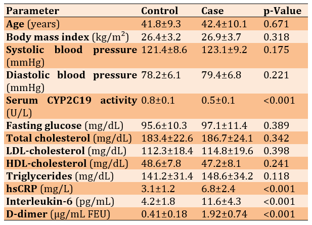
The male-to-female ratio in the case group was 68/37, and in the control group, 70/35 (p=0.822). Cases and controls were similar in age and baseline cardiometabolic parameters (all p>0.05). However, case patients had substantially higher levels of inflammatory and thrombotic markers: hsCRP (p<0.001), IL-6 (p<0.001), and D-dimer (p<0.001). The key finding was a highly significant reduction in CYP2C19 activity in cases compared with controls (p<0.001; Table 1).
Table 1. Comparison of mean values of participants’ baseline demographic, anthropometric, and biochemical characteristics between control (n=105) and case (n=105) groups
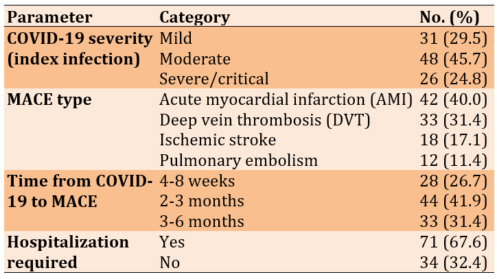
Among 105 cases, AMI was most common (40.0%), followed by DVT (31.4%), ischemic stroke (17.1%), and pulmonary embolism (11.4%). Most MACE occurred 2–3 months after recovery (41.9%), supporting a delayed, inflammation-mediated deterioration rather than an acute mechanism. All cases had no prior cardiovascular disease and no antiplatelet/anticoagulant use at MACE (100% for both), limiting confounding by pre-existing pharmacological risk (Table 2).
Table 2. Frequency of clinical characteristics and major adverse cardiovascular event (MACE) profile of the case group (n=105)
ROC analysis showed that plasma CYP2C19 activity is an excellent diagnostic biomarker for predicting MACE after COVID-19, with an AUC of 0.924 (95% CI: 0.889-0.959; p<0.001). Using an optimal cut-off of 0.648U/L (Youden index) yielded a sensitivity of 86.7% and a specificity of 90.5%, indicating strong single-analyte performance for MACE risk stratification.
MACE risk stratification of post-COVID-19 patients with CYP2C19 activity at or below the threshold of 0.6480 U/L represents a biochemical diagnosis of “Cytokine-Mediated CYP2C19 Insufficiency,” a biochemical state in which enzymatic depletion is severe enough to compromise not only endogenous EET-mediated vascular protection but also, in pharmacologically treated patients, clopidogrel bioactivation.
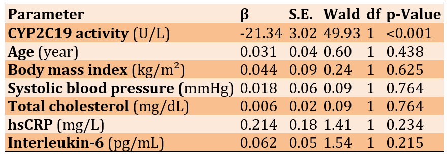
Multivariate logistic regression showed that CYP2C19 activity was the only independent predictor of post-COVID-19 MACE (β=−21.34; p<0.001). In contrast, age, BMI, systolic blood pressure, total cholesterol, hsCRP, and IL-6 were not significant (all p>0.05), suggesting that the association of inflammatory markers with MACE was not independent after adjustment for CYP2C19 activity (Table 3).
Table 3. Multivariate binary logistic regression results
Correlation analysis confirmed that reduced CYP2C19 activity was significantly linked to elevated inflammatory and thrombotic markers—specifically IL-6 (r=-0.512), hsCRP (r=-0.481), and D-dimer (r=-0.443; all p<0.001). The absence of significant correlations with standard cardiometabolic parameters (age, BMI, BP, total cholesterol; all p>0.05) reinforced the specificity of this relationship to the post-COVID-19 thrombo-inflammatory process.
Discussion
This research investigated whether the cytokine-induced inflammatory environment of post-COVID-19 syndrome continuously reduces circulating cytochrome P450 2C19 activity. CYP2C19 activity was markedly and significantly decreased in post-COVID-19 patients who developed MACE, with an effect size that greatly exceeds clinical relevance cutoffs. This reduction in enzymatic activity predicts MACE risk and is independent of other factors after the investigators controlled for eight covariates, including age, sex, BMI, blood pressure, cholesterol, hsCRP, and IL-6. This shows that CYP2C19 insufficiency is not a marker of general inflammatory activity but rather a unique and specific risk factor. The very strong negative correlations between CYP2C19 activity and both IL-6 and hsCRP provided direct biochemical evidence that the post-COVID-19 cytokine environment is linked to enzymatic depletion, thereby completing the proposed mechanistic chain.
The CYP2C19 activity, with an AUC of 0.924, ranks among the highest-performing single-analyte biomarkers described so far for any post-COVID-19 cardiovascular outcome. It even compares favorably with NT-proBNP, used for heart failure risk stratification, and hs-troponin, used to detect AMI [22, 23]. However, unlike the latter biomarkers—which confirm the diagnosis after the event—CYP2C19 activity has the particular advantage of identifying risk prospectively before the onset of MACE.
During and after COVID-19, SARS-CoV-2 interaction with macrophages and the resulting damage to endothelial cells lead to the production of large amounts of cytokines, mostly IL-6, TNF-, and IL-1 [5, 6]. In liver cells, IL-6 receptor interaction activates Janus kinase (JAK) 1/2, which subsequently leads to STAT3 phosphorylation. Once activated, STAT3 translocates to the nucleus, where it hampers the activity of HNF4, the key transcription factor of CYP2C19 gene expression, by competing for the same co-activators such as PGC-1 and SRC-1 [11, 12]. This down-regulation at the transcriptional level leads to reduced synthesis of new CYP2C19 protein. Meanwhile, oxidative stress arising from NOX2 activation and mitochondrial respiratory chain uncoupling results in the direct post-translational inhibition of the existing CYP2C19 protein by haem iron oxidation [13].
The functional impact of CYP2C19 depletion on vascular homeostasis is twofold. First, reduced CYP2C19 epoxygenase activity leads to a fall in the production of epoxyeicosatrienoic acids (EETs) from arachidonic acid. These EETs serve as key internal mediators of vascular protection; thus, a shortage of EETs results in the loss of an essential anti-thrombotic and anti-inflammatory line of defense of the vascular endothelium right at the time when the post-COVID-19 thrombo-inflammatory stimulus is at its strongest. Second, in patients receiving clopidogrel for acute MACE, CYP2C19 deficiency impairs the hepatic conversion of clopidogrel to its active thiol form, thereby causing drug resistance and increased thrombus formation. This phenomenon is well documented in pharmacogenomics for CYP2C19 poor metabolizers [7, 24].
The highly significant negative association between CYP2C19 function and D-dimer links the enzyme's biochemical reduction to the activation of thrombosis. With a decrease in CYP2C19-dependent EET synthesis, the endothelial thrombo-protective barrier is compromised, thereby allowing fibrin cross-linking, as reflected by an increase in D-dimer. This triangular correlation system—IL-6, CYP2C19 repression, and D-dimer increase—constitutes a consistent biochemical story that is compatible with all our primary findings.
Most of the current literature on cardiovascular risk after COVID-19 has linked the main drivers of MACEs to endothelial injury, sustained microthrombi, and ongoing myocardial inflammation [5, 6, 25]. Our work adds a novel aspect; the pharmacokinetic and biochemical effects of cytokine-induced CYP2C19 downregulation on the metabolism of endogenous vascular substrates. This route is independent of, and in addition to, the other pathways discussed previously. It might account for some of the residual cardiovascular risk that post-COVID-19 patients continue to face even after the acute inflammatory phase has resolved and conventional biomarkers have normalized.
Research from non-COVID cohorts has demonstrated that the CYP2C19 poor-metabolizer genotype confers a 30% higher risk for the occurrence of major cardiovascular events among clopidogrel users, along with increased frequencies of in-stent thrombosis [7, 24]. The inflammatory state after COVID-19, which can cause an acquired form of CYP2C19 insufficiency rather than hereditary factors, could confer a similar cardiovascular susceptibility in a large segment of the population, including those with no pharmacogenomic predisposition. Furthermore, this acquired enzymatic phenotype could explain why the risk of MACE after COVID-19 is not perfectly explained by established cardiovascular risk factors or by markers of residual inflammation alone [1, 2].
As this research was conducted in Maysan Province in southern Iraq, it adds more scientific insight. The area is known for its very hot climate, with summer temperatures often exceeding 50°C, high levels of fine dust particles in the air due to sandstorms in different seasons, and a very high level of COVID-19 antibodies in the population. This combination of environmental and infectious oxidative stressors could lead to greater suppression of CYP2C19 than what is observed in populations from temperate climates [16]. Therefore, the results obtained from Maysan represent a community that may be at a significant extreme of cardiometabolic vulnerability after COVID-19 on the spectrum, although the mechanism itself is globally relevant.
We have a clinically actionable single-analyte criterion for risk stratification in post-COVID-19 outpatient follow-up: the validated 0.6480U/L threshold, achieved here in the present cohort with sensitivity 86.7% and specificity 90.5%. The omeprazole OHR method used in this study requires only a single venipuncture at 3 hours after a standard oral omeprazole dose and HPLC-UV quantification, a procedure feasible in any well-equipped clinical biochemistry laboratory, at an estimated reagent cost [14, 15].
The largest number of MACE deaths occurred at 25 months post-recovery; thus, we suggest that CYP2C19 OHR phenotyping be included in structured post-COVID-19 cardiovascular follow-up programs at 8 weeks and 3 months after recovery from moderate-to-severe index illness. Referring post-COVID-19 patients with CYP2C19 activity ≤0.6480U/L for cardiology consultation, non-invasive cardiac workup (ECG, echocardiography, and lower limb Doppler as clinically indicated), and antiplatelet prophylaxis regimens that are independent of CYP2C19 bioactivation (e.g., ticagrelor or prasugrel instead of clopidogrel, since the enzymatic deficit has now been verified) is the way to go.
Using a drug to restore CYP2C19 protein levels by reversing the IL-6/STAT3/HNF4 signaling pathway is a promising approach. The anti–IL-6 receptor antibody tocilizumab, a drug used to treat severe COVID-19, was shown to restore HNF4-dependent CYP gene expression in IL-6-treated hepatocyte models [26]. NAC, as a glutathione precursor, may attenuate the post-translational haem oxidation of CYP2C19. These drugs should be tested in future randomized controlled trials, in which restoration of CYP2C19 enzyme activity is the primary pharmacodynamic endpoint.
The study design is a key strength. Having a matched post-COVID-19 control group, rather than healthy or uninfected comparators, allowed the elimination of infection history as a confounder and directly linked CYP2C19 depletion to MACE risk in the post-COVID-19 cohort. Using the validated, internationally standardized in vivo phenotyping method for CYP2C19: the omeprazole OHR method offers quantitative pharmacokinetic accuracy beyond surrogate serum activity assays. All MACE events were identified using objective, guideline-based criteria (Fourth Universal Definition of MI; imaging-confirmed DVT, stroke, and PE). In addition, event review and adjudication were performed blindly to genotypes. A thorough exclusion framework removed all known pharmacological, genetic, metabolic, and lifestyle confounders of CYP2C19 activity. Besides inflammatory markers, the multivariate regression model with eight covariates provided detailed control of covariates. The three-way correlation network (IL-6, hsCRP, and D-dimer all negatively correlating with CYP2C19) provides multi-analyte mechanistic validation. Finally, the power calculation performed after the study confirmed that the research had sufficient power (99.1%) to detect the observed effect.
Several limitations have been recognized and pointed out explicitly. Because our study was observational and conducted at a single center, causal inference is fundamentally highly limited, whereas external validity would require replicating the study in multiple centers. We did not obtain CYP2C19 genetic polymorphism data; therefore, it was impossible to isolate the suppressive effect of inflammation caused by gene expression deficiency from the constitutive gene defect, which is a crucial point for further research. Traceable CYP2C19 levels starting from the early phase of COVID-19 infection were not accessible. The current cross-sectional enzymatic assays merely depict the state at the time of enrolment, not the pattern of enzymatic reduction. Furthermore, the study sample represents the population of the Maysan governorate. In addition to the high heat and dust, the environmental conditions of this region might limit direct quantitative extrapolation of the threshold value to populations with different background levels of oxidative stress, although the fundamental mechanistic relationships are likely to be the same. In addition, we did not obtain COVID-19 variant samples systematically throughout the recruitment period; therefore, variant-specific differences in cytokine profiles and CYP2C19 suppression levels should be explored in subsequent studies.
Conclusion
Markedly reduced post–COVID-19 CYP2C19 enzymatic activity is a standalone, highly reliable independent biomarker for predicting major adverse cardiac events.
Acknowledgments: The authors would like to express their sincere gratitude to the University of Manara for its continuous support and encouragement throughout this study.
Ethical Permissions: Ethical approval for this study was obtained from the Institutional Ethics Committee of the University of Mustansiriyah, Iraq (Approval Reference No.: 2022/146C, dated January 20, 2022).
Conflicts of Interest: The authors declared no conflicts of interest.
Authors' Contribution: Jasim AK (First Author), Methodologist/Main Researcher/Statistical Analyst (60%); Hamadi HS (Second Author), Introduction Writer/Assistant Researcher/Discussion Writer (40%)
Funding/Support: The authors declared that this research did not receive any specific grants from public funding agencies.
Infection with SARS-CoV-2 leads to a multisystem illness that, among other effects, can cause long-lasting changes in the heart after the infection has resolved. Researchers who have studied very large groups of people have found that those who have had COVID-19 are at a much higher risk of developing new heart disease within a year after they recover, such as heart attack, stroke, heart failure, and blood clots, even if they did not have any heart problems before [1, 2]. This set of heart-related problems after the illness, along with many other symptoms, makes up what have been termed Post-Acute Sequelae of SARS-CoV-2 infection (PASC), or Long COVID, and it is estimated that 10 to 30% of those who have had COVID-19 are dealing with it [3, 4].
A variety of pathobiological processes contribute to sustained cardiovascular risk in post-COVID-19. These include persistently impaired endothelial function, overactive platelets, microvascular thromboinflammation, autonomic dysregulation, and continuous high levels of inflammatory cytokines [5, 6]. However, an important component of the risk puzzle—one that has received little attention so far—is how the cytokine environment after COVID-19 influences the hepatic cytochrome P450 (CYP) enzyme system, the main metabolic system in the body, which, among other functions, handles the processing of both externally and internally produced vasoactive substances.
Cytochrome P450 2C19 (CYP2C19) is considered one of the most crucial CYP isoforms clinically in the field of cardiovascular medication. It is the enzyme that, in the liver, converts the antiplatelet prodrug clopidogrel into its active thiol metabolite [7]. It also metabolizes arachidonic acid into epoxyeicosatrienoic acids (EETs)—which are potent vasodilators and have antithrombotic properties [8]—and it is involved in the biotransformation of several endogenous vasoactive substances, including thromboxane precursors. Besides its pharmacological function, CYP2C19-derived EETs also directly protect the vascular endothelium by blocking NF-B–triggered inflammation, reducing leukocyte adhesion, and maintaining the balance of fibrinolysis [9, 10].
Significant transcriptional downregulation of CYP2C19 occurs during systemic inflammation. Pro-inflammatory cytokines IL-6 and TNF—both of which are present at significantly high levels during and after COVID-19 infection—lead to the activation of STAT3 and NF-B signaling pathways within hepatocytes, which in turn causes phosphorylation-dependent inhibition of hepatocyte nuclear factor 4 (HNF4), the main transcriptional regulator of CYP2C19 [11, 12]. At the same time, a continuous oxidative stress environment resulting from COVID-19-induced NADPH oxidase activation will cause post-translational heme degradation of the CYP2C19 protein via formation of the compound I ferryl intermediate [13]. Therefore, the combined transcriptional and post-translational repression of CYP2C19 by the post-COVID-19 inflammatory milieu provides a fully plausible biological explanation for the progressive enzymatic deficiency that can persist for weeks to months after viral elimination.
Although this mechanistic plausibility cannot be ruled out, to date, no study has prospectively quantified CYP2C19 enzymatic activity in post-COVID-19 patients and simultaneously assessed the relationship between CYP2C19 enzymatic activity and major adverse cardiovascular events (MACEs) using a matched post-COVID-19 control design [14, 15]. With its demographic and environmental characteristics—including high ambient temperatures, dust-related oxidative stress exposure, and, in addition to the historically high COVID-19 case burden—this population is particularly informative for studying post-COVID-19 cardiometabolic susceptibility [16]. Decreased CYP2C19 enzymatic activity is an associated factor for cardiovascular risk, even after adjusting for well-known risk factors and inflammatory markers [17].
This study aimed to investigate whether CYP2C19 enzymatic activity decreases substantially in post-COVID-19 individuals who develop MACEs compared with those who remain event-free.
Instrument and Methods
This prospective, single-center, case-control study was conducted at the Maysan Center for Heart Diseases and Surgery, Maysan Health Directorate, Amarah, Maysan Province, Iraq, from October 2022 to November 2024. This Center is the main tertiary referral center for Maysan governorate, with a registered population of about 1.1 million. The hospital’s cardiology department manages the full spectrum of acute and chronic cardiovascular diseases in the region, including all post-COVID-19 cardiovascular cases referred from the province’s primary and secondary care centers.
Altogether, 210 adult participants were recruited from the outpatient cardiology and post-COVID-19 follow-up clinics of the Maysan Center for Heart Diseases and Surgery and divided into two groups. Both groups consisted of individuals with PCR-confirmed COVID-19 infections during the time of study enrollment.
The case group (n=105) included adults who developed a new MACE (objectively confirmed) within six months of COVID-19 recovery, defined as a heart attack, deep vein thrombosis, ischemic stroke, or pulmonary embolism. The diagnosis of heart attack was based on the Fourth Universal Definition criteria (cardiac troponin rise/fall plus symptoms or ECG changes indicative of myocardial ischemia). Deep vein thrombosis (DVT) was detected by Duplex Doppler ultrasound. Ischemic stroke was based on clinical assessment and CT/MRI imaging. Pulmonary embolism (PE) was diagnosed through CT pulmonary angiography [19].
The control group (n=105) comprised individuals who had recovered from COVID-19 and visited the same institutional follow-up clinic during the same calendar period as the cases. Controls were matched to cases by age, sex, and BMI (2.5kg/m). Control participants included not only those who completed a structured six-month cardiovascular surveillance program but also those who did not develop any MACE. Recruitment of controls was prospective and concurrent with case identification, ensuring temporal comparability of post-infection inflammatory status.
A matched post-COVID-19 versus post-COVID-19 design such as this one excludes the confounding effect of infection history and highlights enzymatic depletion as a primary factor underlying adverse cardiovascular outcomes in the post-COVID-19 population. All patients admitted to the cardiac hospital during the research who met the inclusion criteria were enrolled. Each eligible case was recorded individually. Consequently, the sample size was not formally determined, because all available cases during the research were included.
Participants were eligible only if they were adults (>18 years) of either gender with a documented history of PCR-confirmed COVID-19 (controls were eligible if they attended follow-up after recovery) and had no prior diagnosis of cardiovascular disease. Participants were also excluded if they had conditions known to alter CYP2C19 expression/activity independent of COVID-19 (e.g., cirrhosis/major liver dysfunction with ALT or AST>3× upper normal limit, chronic kidney disease with eGFR <45mL/min/1.73m², active cancer, or type 1 diabetes), current or recent (last 30 days) use of CYP2C19 inhibitors (e.g., fluconazole, fluvoxamine, ticlopidine, and chronic omeprazole) or potent CYP2C19 inducers (e.g., rifampicin, carbamazepine, St John’s Wort), confirmed/suspected hereditary thrombophilia (e.g., factor V Leiden, prothrombin G20210A, protein C/S or antithrombin III deficiency), active smoking (excluded due to smoking-induced effects on multiple CYP enzymes including CYP2C19) [20], pregnancy/lactation, or BMI>35kg/m².
All biochemical analyses were performed in the hospital’s accredited clinical biochemistry laboratory under standardized pre-analytical conditions, and the laboratory staff was not aware of the participants’ group allocations.
The study was conducted in accordance with the ethical standards set forth in the 1964 Declaration of Helsinki and its six amendments through 2013 [18]. Each potential subject was invited to a clear and detailed informed-consent discussion, in which the main points of the study were presented, the types and quantities of biological specimens were described, and it was explained that the data would be fully anonymized. In addition, the participants’ rights to withdraw at any time without their care being affected were explained in detail. Subsequently, written informed consent was obtained from each participant before the initiation of any study-related procedures. In line with national Iraqi health data protection rules, personal and clinical data were anonymized and securely stored.
CYP2C19 phenotyping was performed using a validated method based on the omeprazole hydroxylation ratio (OHR). This measure determines the plasma ratio of 5-hydroxy omeprazole to omeprazole after a standard oral dose of omeprazole—the internationally recognized reference method for non-invasive in vivo CYP2C19 phenotyping [14, 15]. Each participant received a single oral dose of omeprazole 20mg in the fasting state. Venous blood samples (10mL) were collected exactly 3 hours after dosing into EDTA-containing vacutainers. Plasma was obtained by centrifuging the tubes at 2,500g for 10 minutes at 4°C and then freezing the samples at -80°C until analysis, which was conducted in batches within 4 weeks of collection.
The concentrations of omeprazole and 5-hydroxyomeprazole in plasma were measured using a validated high-performance liquid chromatography method with ultraviolet detection (HPLC-UV) at a wavelength of 302nm. A C18 reverse-phase analytical column (Zorbax Eclipse XDB-C18, 4.6×150mm, 5µm) was used with isocratic elution and ammonium acetate/acetonitrile as the mobile phase. Calibration standard curves for both analytes were prepared from certified reference standards (Sigma-Aldrich, USA) in the concentration range 5-2000ng/mL. The OHR was calculated as the molar ratio of 5-hydroxy omeprazole to omeprazole. To maintain consistency, the values were reported in metabolic activity units (U/L). The intra-assay coefficient of variation was <4.2%, and the inter-assay coefficient of variation was <5.8% for all batches. The CYP2C19 poor-metabolizer phenotype was identified as OHR<0.8 according to the established reference ranges [21].
All participants had venous blood drawn after standardized fasting for the following tests: complete blood count with differential, fasting plasma glucose, full lipid profile (total cholesterol, LDL-C, HDL-C, triglycerides), renal function panel (creatinine, urea, eGFR), liver enzyme panel (ALT, AST, ALP, GGT), serum high-sensitivity C-reactive protein (hsCRP; immunoturbidimetric method), plasma interleukin-6 (IL-6; enzyme-linked immunosorbent assay, ELISA; Abcam ab178013), and plasma D-dimer (immunoturbidimetric latex agglutination; Siemens INNOVANCE D-Dimer). All the tests were conducted in the hospital’s accredited biochemistry laboratory using calibrated automated analyzers.
Statistical analyses were conducted using SPSS 26 and GraphPad Prism 10. The Shapiro-Wilk test indicated that all continuous parameters were normally distributed (all p>0.05). Independent-samples Student’s t-test was used to evaluate between-group differences, and Levene’s test was used to verify homogeneity of variance. Categorical parameters were compared using the Chi-square test. The magnitude of effect for continuous comparisons was determined by Cohen’s d.
Multivariate binary logistic regression was used to determine independent predictors of MACE, and pre-specified covariates, including CYP2C19 activity (U/L), age (years), sex (binary), BMI (kg/m2), systolic blood pressure (mmHg), total cholesterol (mg/dL), hsCRP (mg/L), and IL-6 (pg/mL) were included. The significance of predictors was determined using the Wald statistic. Receiver operating characteristic (ROC) curve analysis was conducted to determine the best diagnostic threshold by maximizing the Youden index (J=sensitivity+specificity−1). Pearson correlation coefficients were used to measure the linear relationships between CYP2C19 activity and continuous biochemical and biometric parameters.
A post-hoc power analysis with G*Power 3.1 showed that n=210 provides 99.1% power to detect the observed effect size (Cohen’s d=2.98) with a two-tailed α=0.05.
Findings
The male-to-female ratio in the case group was 68/37, and in the control group, 70/35 (p=0.822). Cases and controls were similar in age and baseline cardiometabolic parameters (all p>0.05). However, case patients had substantially higher levels of inflammatory and thrombotic markers: hsCRP (p<0.001), IL-6 (p<0.001), and D-dimer (p<0.001). The key finding was a highly significant reduction in CYP2C19 activity in cases compared with controls (p<0.001; Table 1).
Table 1. Comparison of mean values of participants’ baseline demographic, anthropometric, and biochemical characteristics between control (n=105) and case (n=105) groups
Among 105 cases, AMI was most common (40.0%), followed by DVT (31.4%), ischemic stroke (17.1%), and pulmonary embolism (11.4%). Most MACE occurred 2–3 months after recovery (41.9%), supporting a delayed, inflammation-mediated deterioration rather than an acute mechanism. All cases had no prior cardiovascular disease and no antiplatelet/anticoagulant use at MACE (100% for both), limiting confounding by pre-existing pharmacological risk (Table 2).
Table 2. Frequency of clinical characteristics and major adverse cardiovascular event (MACE) profile of the case group (n=105)
ROC analysis showed that plasma CYP2C19 activity is an excellent diagnostic biomarker for predicting MACE after COVID-19, with an AUC of 0.924 (95% CI: 0.889-0.959; p<0.001). Using an optimal cut-off of 0.648U/L (Youden index) yielded a sensitivity of 86.7% and a specificity of 90.5%, indicating strong single-analyte performance for MACE risk stratification.
MACE risk stratification of post-COVID-19 patients with CYP2C19 activity at or below the threshold of 0.6480 U/L represents a biochemical diagnosis of “Cytokine-Mediated CYP2C19 Insufficiency,” a biochemical state in which enzymatic depletion is severe enough to compromise not only endogenous EET-mediated vascular protection but also, in pharmacologically treated patients, clopidogrel bioactivation.
Multivariate logistic regression showed that CYP2C19 activity was the only independent predictor of post-COVID-19 MACE (β=−21.34; p<0.001). In contrast, age, BMI, systolic blood pressure, total cholesterol, hsCRP, and IL-6 were not significant (all p>0.05), suggesting that the association of inflammatory markers with MACE was not independent after adjustment for CYP2C19 activity (Table 3).
Table 3. Multivariate binary logistic regression results
Correlation analysis confirmed that reduced CYP2C19 activity was significantly linked to elevated inflammatory and thrombotic markers—specifically IL-6 (r=-0.512), hsCRP (r=-0.481), and D-dimer (r=-0.443; all p<0.001). The absence of significant correlations with standard cardiometabolic parameters (age, BMI, BP, total cholesterol; all p>0.05) reinforced the specificity of this relationship to the post-COVID-19 thrombo-inflammatory process.
Discussion
This research investigated whether the cytokine-induced inflammatory environment of post-COVID-19 syndrome continuously reduces circulating cytochrome P450 2C19 activity. CYP2C19 activity was markedly and significantly decreased in post-COVID-19 patients who developed MACE, with an effect size that greatly exceeds clinical relevance cutoffs. This reduction in enzymatic activity predicts MACE risk and is independent of other factors after the investigators controlled for eight covariates, including age, sex, BMI, blood pressure, cholesterol, hsCRP, and IL-6. This shows that CYP2C19 insufficiency is not a marker of general inflammatory activity but rather a unique and specific risk factor. The very strong negative correlations between CYP2C19 activity and both IL-6 and hsCRP provided direct biochemical evidence that the post-COVID-19 cytokine environment is linked to enzymatic depletion, thereby completing the proposed mechanistic chain.
The CYP2C19 activity, with an AUC of 0.924, ranks among the highest-performing single-analyte biomarkers described so far for any post-COVID-19 cardiovascular outcome. It even compares favorably with NT-proBNP, used for heart failure risk stratification, and hs-troponin, used to detect AMI [22, 23]. However, unlike the latter biomarkers—which confirm the diagnosis after the event—CYP2C19 activity has the particular advantage of identifying risk prospectively before the onset of MACE.
During and after COVID-19, SARS-CoV-2 interaction with macrophages and the resulting damage to endothelial cells lead to the production of large amounts of cytokines, mostly IL-6, TNF-, and IL-1 [5, 6]. In liver cells, IL-6 receptor interaction activates Janus kinase (JAK) 1/2, which subsequently leads to STAT3 phosphorylation. Once activated, STAT3 translocates to the nucleus, where it hampers the activity of HNF4, the key transcription factor of CYP2C19 gene expression, by competing for the same co-activators such as PGC-1 and SRC-1 [11, 12]. This down-regulation at the transcriptional level leads to reduced synthesis of new CYP2C19 protein. Meanwhile, oxidative stress arising from NOX2 activation and mitochondrial respiratory chain uncoupling results in the direct post-translational inhibition of the existing CYP2C19 protein by haem iron oxidation [13].
The functional impact of CYP2C19 depletion on vascular homeostasis is twofold. First, reduced CYP2C19 epoxygenase activity leads to a fall in the production of epoxyeicosatrienoic acids (EETs) from arachidonic acid. These EETs serve as key internal mediators of vascular protection; thus, a shortage of EETs results in the loss of an essential anti-thrombotic and anti-inflammatory line of defense of the vascular endothelium right at the time when the post-COVID-19 thrombo-inflammatory stimulus is at its strongest. Second, in patients receiving clopidogrel for acute MACE, CYP2C19 deficiency impairs the hepatic conversion of clopidogrel to its active thiol form, thereby causing drug resistance and increased thrombus formation. This phenomenon is well documented in pharmacogenomics for CYP2C19 poor metabolizers [7, 24].
The highly significant negative association between CYP2C19 function and D-dimer links the enzyme's biochemical reduction to the activation of thrombosis. With a decrease in CYP2C19-dependent EET synthesis, the endothelial thrombo-protective barrier is compromised, thereby allowing fibrin cross-linking, as reflected by an increase in D-dimer. This triangular correlation system—IL-6, CYP2C19 repression, and D-dimer increase—constitutes a consistent biochemical story that is compatible with all our primary findings.
Most of the current literature on cardiovascular risk after COVID-19 has linked the main drivers of MACEs to endothelial injury, sustained microthrombi, and ongoing myocardial inflammation [5, 6, 25]. Our work adds a novel aspect; the pharmacokinetic and biochemical effects of cytokine-induced CYP2C19 downregulation on the metabolism of endogenous vascular substrates. This route is independent of, and in addition to, the other pathways discussed previously. It might account for some of the residual cardiovascular risk that post-COVID-19 patients continue to face even after the acute inflammatory phase has resolved and conventional biomarkers have normalized.
Research from non-COVID cohorts has demonstrated that the CYP2C19 poor-metabolizer genotype confers a 30% higher risk for the occurrence of major cardiovascular events among clopidogrel users, along with increased frequencies of in-stent thrombosis [7, 24]. The inflammatory state after COVID-19, which can cause an acquired form of CYP2C19 insufficiency rather than hereditary factors, could confer a similar cardiovascular susceptibility in a large segment of the population, including those with no pharmacogenomic predisposition. Furthermore, this acquired enzymatic phenotype could explain why the risk of MACE after COVID-19 is not perfectly explained by established cardiovascular risk factors or by markers of residual inflammation alone [1, 2].
As this research was conducted in Maysan Province in southern Iraq, it adds more scientific insight. The area is known for its very hot climate, with summer temperatures often exceeding 50°C, high levels of fine dust particles in the air due to sandstorms in different seasons, and a very high level of COVID-19 antibodies in the population. This combination of environmental and infectious oxidative stressors could lead to greater suppression of CYP2C19 than what is observed in populations from temperate climates [16]. Therefore, the results obtained from Maysan represent a community that may be at a significant extreme of cardiometabolic vulnerability after COVID-19 on the spectrum, although the mechanism itself is globally relevant.
We have a clinically actionable single-analyte criterion for risk stratification in post-COVID-19 outpatient follow-up: the validated 0.6480U/L threshold, achieved here in the present cohort with sensitivity 86.7% and specificity 90.5%. The omeprazole OHR method used in this study requires only a single venipuncture at 3 hours after a standard oral omeprazole dose and HPLC-UV quantification, a procedure feasible in any well-equipped clinical biochemistry laboratory, at an estimated reagent cost [14, 15].
The largest number of MACE deaths occurred at 25 months post-recovery; thus, we suggest that CYP2C19 OHR phenotyping be included in structured post-COVID-19 cardiovascular follow-up programs at 8 weeks and 3 months after recovery from moderate-to-severe index illness. Referring post-COVID-19 patients with CYP2C19 activity ≤0.6480U/L for cardiology consultation, non-invasive cardiac workup (ECG, echocardiography, and lower limb Doppler as clinically indicated), and antiplatelet prophylaxis regimens that are independent of CYP2C19 bioactivation (e.g., ticagrelor or prasugrel instead of clopidogrel, since the enzymatic deficit has now been verified) is the way to go.
Using a drug to restore CYP2C19 protein levels by reversing the IL-6/STAT3/HNF4 signaling pathway is a promising approach. The anti–IL-6 receptor antibody tocilizumab, a drug used to treat severe COVID-19, was shown to restore HNF4-dependent CYP gene expression in IL-6-treated hepatocyte models [26]. NAC, as a glutathione precursor, may attenuate the post-translational haem oxidation of CYP2C19. These drugs should be tested in future randomized controlled trials, in which restoration of CYP2C19 enzyme activity is the primary pharmacodynamic endpoint.
The study design is a key strength. Having a matched post-COVID-19 control group, rather than healthy or uninfected comparators, allowed the elimination of infection history as a confounder and directly linked CYP2C19 depletion to MACE risk in the post-COVID-19 cohort. Using the validated, internationally standardized in vivo phenotyping method for CYP2C19: the omeprazole OHR method offers quantitative pharmacokinetic accuracy beyond surrogate serum activity assays. All MACE events were identified using objective, guideline-based criteria (Fourth Universal Definition of MI; imaging-confirmed DVT, stroke, and PE). In addition, event review and adjudication were performed blindly to genotypes. A thorough exclusion framework removed all known pharmacological, genetic, metabolic, and lifestyle confounders of CYP2C19 activity. Besides inflammatory markers, the multivariate regression model with eight covariates provided detailed control of covariates. The three-way correlation network (IL-6, hsCRP, and D-dimer all negatively correlating with CYP2C19) provides multi-analyte mechanistic validation. Finally, the power calculation performed after the study confirmed that the research had sufficient power (99.1%) to detect the observed effect.
Several limitations have been recognized and pointed out explicitly. Because our study was observational and conducted at a single center, causal inference is fundamentally highly limited, whereas external validity would require replicating the study in multiple centers. We did not obtain CYP2C19 genetic polymorphism data; therefore, it was impossible to isolate the suppressive effect of inflammation caused by gene expression deficiency from the constitutive gene defect, which is a crucial point for further research. Traceable CYP2C19 levels starting from the early phase of COVID-19 infection were not accessible. The current cross-sectional enzymatic assays merely depict the state at the time of enrolment, not the pattern of enzymatic reduction. Furthermore, the study sample represents the population of the Maysan governorate. In addition to the high heat and dust, the environmental conditions of this region might limit direct quantitative extrapolation of the threshold value to populations with different background levels of oxidative stress, although the fundamental mechanistic relationships are likely to be the same. In addition, we did not obtain COVID-19 variant samples systematically throughout the recruitment period; therefore, variant-specific differences in cytokine profiles and CYP2C19 suppression levels should be explored in subsequent studies.
Conclusion
Markedly reduced post–COVID-19 CYP2C19 enzymatic activity is a standalone, highly reliable independent biomarker for predicting major adverse cardiac events.
Acknowledgments: The authors would like to express their sincere gratitude to the University of Manara for its continuous support and encouragement throughout this study.
Ethical Permissions: Ethical approval for this study was obtained from the Institutional Ethics Committee of the University of Mustansiriyah, Iraq (Approval Reference No.: 2022/146C, dated January 20, 2022).
Conflicts of Interest: The authors declared no conflicts of interest.
Authors' Contribution: Jasim AK (First Author), Methodologist/Main Researcher/Statistical Analyst (60%); Hamadi HS (Second Author), Introduction Writer/Assistant Researcher/Discussion Writer (40%)
Funding/Support: The authors declared that this research did not receive any specific grants from public funding agencies.
Article Type: Original Research |
| Rights and permissions | |
 |
This work is licensed under a Creative Commons Attribution-NonCommercial 4.0 International License. |